

Researchers Develop Method to Study Brain Connectivity, Functionality
STANFORD, Calif. — Researchers at Stanford University have developed a research method that enables them to take a much more detailed look at the brain processes connected to some neurological and mental disorders.
What researcher Dr. Sergiu Pasca and colleagues at Stanford did was demonstrate they could grow human cortical organoids in a lab culture and then insert them into developing rodent brains to see how they integrate and function over time.
The findings suggest that transplanted organoids may offer a powerful tool for investigating the processes associated with disease development.
The study, funded by the National Institute of Mental Health, part of the National Institutes of Health, appears in the journal Nature.
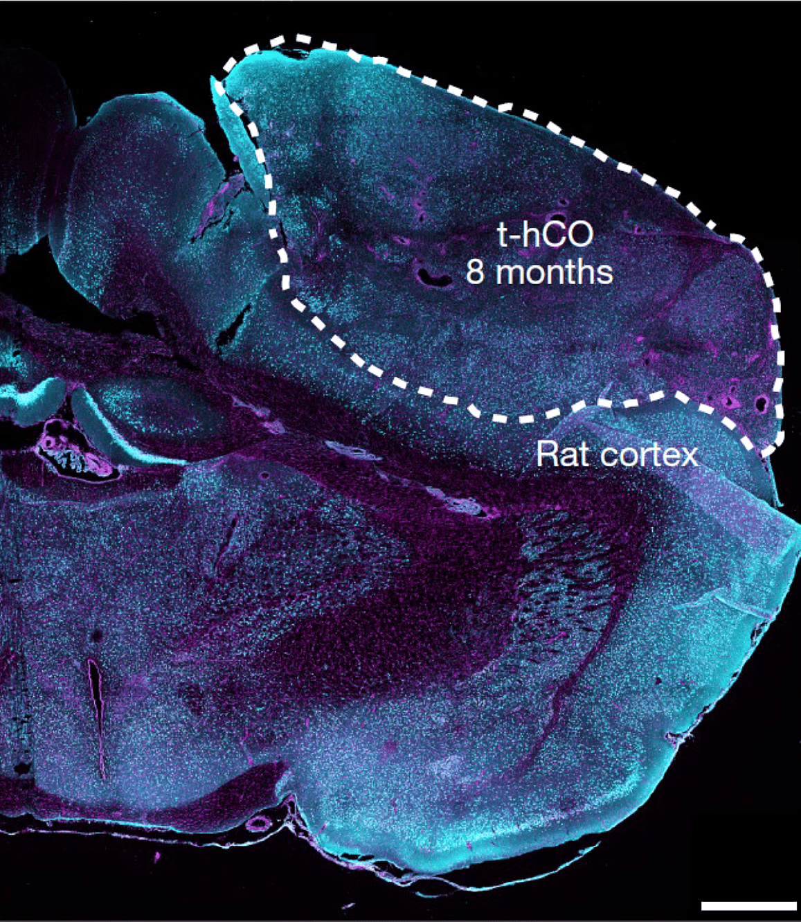
In this study, the team of researchers advanced the use of brain organoids for research by transplanting an intact human cortical organoid into a developing rat brain. This technique creates a unit of human tissue that can be examined and manipulated.
The researchers used methods previously pioneered in the Pasca lab to create cortical organoids using human-induced pluripotent stem cells — cells derived from adult skin cells that have been reprogrammed into an immature stem-cell-like state. They then implanted these organoids onto the rat primary somatosensory cortex, a part of the brain involved in processing sensation.
The researchers did not detect any motor or memory abnormalities or abnormalities in brain activity in the rats that received the transplanted organoid. Blood vessels from the rat brain successfully supported the implanted tissue, which grew over time.
Structurally and functionally, after seven to eight months of growth, the transplanted brain organoid resembled neurons from human brain tissue more than human organoids maintained in cell culture. The fact that the transplanted organoids mirrored the structural and functional features of human cortical neurons led the researchers to wonder if they could use transplanted organoids to examine aspects of human disease processes.
To examine this, the researchers generated cortical organoids with cells from three participants with a rare genetic disorder associated with autism and epilepsy called Timothy Syndrome and three participants without any known diseases and implanted them onto the rat brain.
Both types of organoids integrated into the rat somatosensory cortex, but organoids derived from Timothy Syndrome patients displayed structural differences. These structural differences did not appear in organoids that were created from the cells of patients with Timothy Syndrome and maintained in cell culture.
“These experiments suggest that this novel approach can capture processes that go beyond what we can detect with current in vitro models,” said Pasca. “This is important because many of the changes that cause psychiatric disease are likely subtle differences at the circuit level.”
Dan can be reached at [email protected] and at https://twitter.com/DanMcCue.